How to Convert Medical DICOM Scans to a High-Resolution PDF
Summary
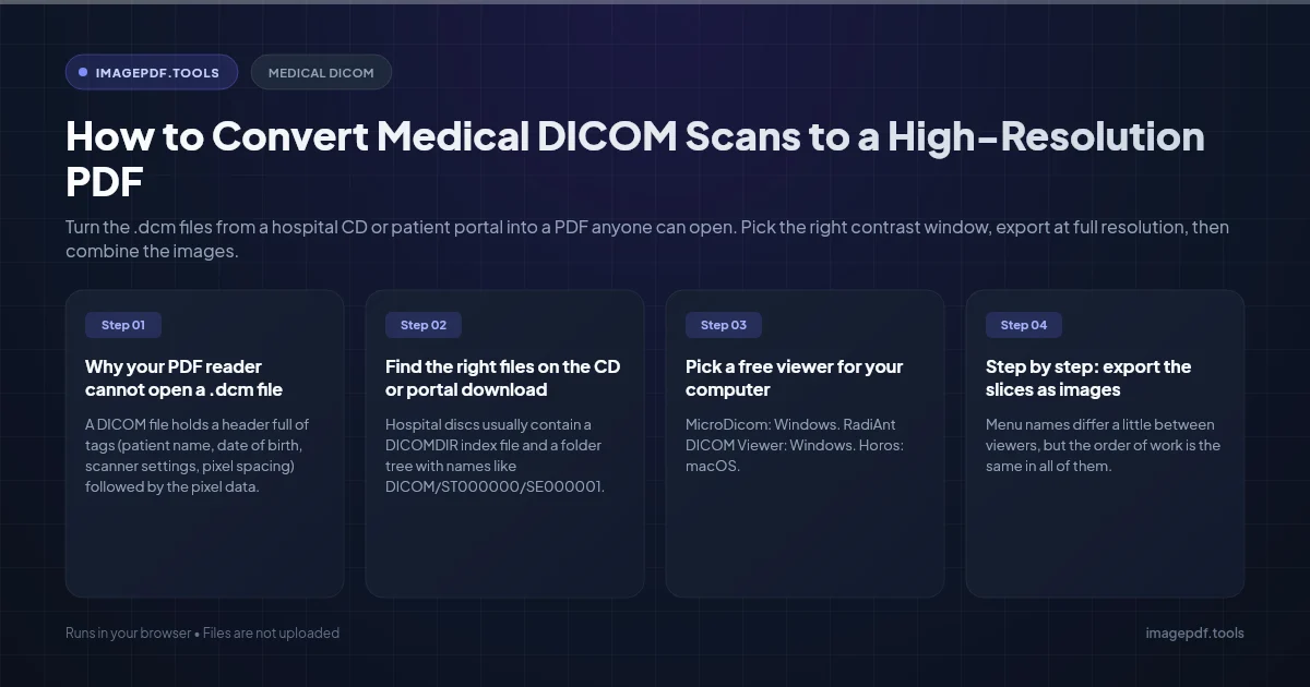
Turn the .dcm files from a hospital CD or patient portal into a PDF anyone can open. Pick the right contrast window, export at full resolution, then combine the images.
There is no one-click DICOM to PDF button in Word, Preview or a phone's file app, because a DICOM file is not an ordinary picture. The reliable route has two stages. First, open the scan in a free DICOM viewer, set the contrast you want to show, and export the slices you care about as PNG or JPEG images. Second, put those images into one PDF.
Before you start, be clear about what the PDF is for. It is a good way to show a family member, a physiotherapist or an insurer what a scan looks like. It is not a replacement for the original study. A radiologist or surgeon giving a second opinion will want the DICOM files themselves, because a PDF freezes one contrast setting and drops the measurements the viewer software relies on.
Why your PDF reader cannot open a .dcm file
A DICOM file holds a header full of tags (patient name, date of birth, scanner settings, pixel spacing) followed by the pixel data. That pixel data usually has 12 to 16 bits per pixel, which means thousands of shades of grey. A normal screen shows 256. Something has to decide which slice of that range becomes black, which becomes white, and what happens in between.
That decision is called windowing. On a CT scan, a lung window and a bone window applied to the same slice look like two different pictures. A generic image converter that ignores the window tags in the header will often give you a frame that is almost entirely white, black or flat grey. A proper DICOM viewer reads those tags and lets you adjust them with the mouse.
There is one more trap. Some X-ray files use a photometric setting called MONOCHROME1, where low values are meant to display as white. A tool that does not check for it produces an inverted, negative-looking image.
Find the right files on the CD or portal download
Hospital discs usually contain a DICOMDIR index file and a folder tree with names like DICOM/ST000000/SE000001. The image files inside often have no extension at all. Do not rename them. Open the DICOMDIR, or the whole folder, from inside the viewer so it can group the files into series.
Many discs also include a small viewer that runs from the disc on Windows, and a PDF or text copy of the radiologist's report. Check for the report first. If what you need is the written finding, it may already be in a PDF.
Pick a free viewer for your computer
| Viewer | Platform | Cost | What it exports |
|---|---|---|---|
| MicroDicom | Windows | Free for non-commercial use | JPEG, PNG, TIFF, BMP, GIF images and video; also has an anonymizer |
| RadiAnt DICOM Viewer | Windows | Paid licence | JPEG or BMP images and WMV video; PDF only through a PDF printer |
| Horos | macOS | Free, open source | JPEG and TIFF images, movies |
| Weasis | Windows, macOS, Linux | Free, open source | Images of the current view |
Step by step: export the slices as images
Menu names differ a little between viewers, but the order of work is the same in all of them.
- 1.Open the study from the DICOMDIR file or the folder, then pick the series you want. A CT often has several: thin slices, thick slices, and reconstructions in other planes.
- 2.Scroll to the slices that show the finding. If you have the report, it often names the series and image number, for example "series 3, image 112".
- 3.Set the window. Most viewers have presets such as lung, bone, brain or abdomen for CT. For MRI and X-ray, drag with the window/level tool until the anatomy is clear.
- 4.Hide or keep the text overlay. The corner text usually shows the patient's name and date of birth. Turn it off if the PDF is going to anyone outside your care team.
- 5.Export as PNG if the viewer offers it, otherwise JPEG at the highest quality. In MicroDicom this is under File, Export. Export only the slices you need, not all 400 of a CT series.
- 6.Choose actual size or 100 percent. CT slices are usually 512 by 512 pixels and an X-ray can be over 2,000 pixels on a side. Scaling up adds file size, not detail.
Combine the images into one PDF
Once you have a folder of PNG or JPEG files, any image to PDF tool will finish the job. Our Image to PDF tool runs in your browser, so the scans are not uploaded anywhere. Drop the images in, drag them into order, and choose A4, Letter, or Fit so each page matches its image. JPEG and PNG files are placed into the PDF as they are, without being compressed again, so the grey levels you exported stay intact.
Name the files with the slice number first (for example CT-chest-112.png) so they sort into the right order before you start dragging.
Removing the overlay text does not clean the image of everything. Some X-rays and ultrasound images have the patient's name burned into the pixels themselves. Look at every page at full size before you send the PDF.
For many studies: a short Python script
If you work with research data and need to convert many files the same way, pydicom and Pillow can apply the stored window and write a multi-page PDF. This uses pydicom 3, where the lookup table helpers live in pydicom.pixels.
from pathlib import Path
import numpy as np
import pydicom
from pydicom.pixels import apply_modality_lut, apply_voi_lut
from PIL import Image

pages = []
for path in sorted(Path("series").glob("*.dcm")):
    ds = pydicom.dcmread(path)
    arr = apply_modality_lut(ds.pixel_array, ds)  # stored values to real units, e.g. Hounsfield
    arr = apply_voi_lut(arr, ds).astype(float)    # window center and width from the header
    arr = (arr - arr.min()) / max(arr.max() - arr.min(), 1) * 255
    if ds.PhotometricInterpretation == "MONOCHROME1":
        arr = 255 - arr                           # low values should be white
    pages.append(Image.fromarray(arr.astype(np.uint8)))

pages[0].save("series.pdf", save_all=True, append_images=pages[1:])
Two things trip people up here. Compressed DICOM files (JPEG Lossless and JPEG 2000 are common) need an extra decoder package such as pylibjpeg before pixel_array will work. And if a file has no window tags, the script stretches the full range, which can look washed out. Set your own center and width in that case.
Privacy: who you are sending it to matters
You can share your own medical images with whoever you like. The rules that bind hospitals and clinics in the US are about how they handle your records, not about what you do with your copy. Still, a scan with your name, date of birth and hospital number is sensitive. Turn off overlays, use a viewer's anonymize feature when you have one, and avoid uploading the raw DICOM files to random online converters, since the header carries far more personal detail than the picture shows.
Frequently asked questions
Can a doctor diagnose from a PDF of my scan?
My exported image is completely white or black. What went wrong?
Should I export PNG or JPEG?
Can I convert a whole CT series of several hundred slices?
Can I open DICOM files on a phone?
Sources & references
This article was researched and written by Nikola, drawing on the following primary sources and documentation:
Ready to try it?
All tools run entirely in your browser, no uploads, no account required.
Image to PDF